
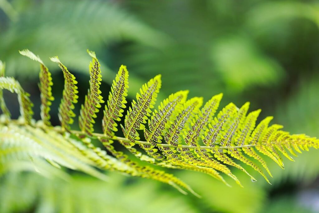
Mushroom enthusiasts often rely on visual characteristics to identify species, but this method can be misleading due to variability in color, shape, and size. Spore print analysis is a more accurate technique for mushroom identification, used by researchers, mycologists, and forensic experts alike. When done correctly, spore prints can reveal crucial information about a specimen’s identity. To collect a reliable spore print, you’ll need a few simple tools: a microscope slide, a petri dish, a razor blade, and a clean surface to work on. In this article, we’ll walk through the step-by-step process of collecting and interpreting spore prints, covering equipment selection, techniques for getting a clear print, common issues that can arise, and how to troubleshoot them. By following these instructions, you’ll be able to accurately collect and analyze spore prints for mushroom identification, research, or forensic analysis.
Understanding the Importance of Spore Prints
To accurately identify a mushroom, it’s crucial to understand why spore prints are essential and how they help distinguish between different species. This knowledge will guide you in taking accurate spore print samples.
What Are Spore Prints Used For?
In mycology, spore prints are a crucial tool for identifying and classifying mushroom species. They provide a visual representation of the spores’ color, shape, size, and arrangement, which are critical characteristics for distinguishing between species. By analyzing spore prints, researchers can gain insights into the reproductive habits and ecological niches of various fungi.
Spore prints also play a significant role in research purposes, such as studying fungal evolution, ecology, and biology. Scientists use spores to investigate the relationships between different species, understand their interactions with their environments, and explore potential applications for biotechnology. For example, researchers have used spore prints to study the mycorrhizal networks of forest ecosystems, revealing complex relationships between fungi and tree roots.
In addition to identification and research purposes, spore prints can be used in forensic analysis to help solve crimes related to mushroom-based food adulteration or contamination. By comparing the characteristics of a suspect’s spores with known reference collections, investigators can determine the origin and authenticity of a mushroom sample.
Preparing for Spore Print Collection
To collect high-quality spore prints, you’ll need a few essential pieces of equipment and some specific materials. A microscope is crucial for examining the spores’ color, shape, and size, which are vital characteristics for identification. You can use a stereo microscope or a compound microscope, depending on your expertise and available budget.
In addition to a microscope, you’ll also need tweezers for handling the mushroom caps without damaging them. It’s essential to choose sterile tweezers to prevent contamination and ensure accurate results. A sterile surface is also necessary for preparing the spore print; this can be achieved by using a petri dish or a glass slide.
When preparing your equipment, make sure it’s clean and free of any residue. You’ll also need a few other materials, such as a small brush for gently removing excess spores from the cap and a paper towel for blotting the mushroom surface. By having these tools at hand, you can collect high-quality spore prints that will aid in accurate identification.
Selecting the Right Specimen
When selecting a specimen for spore print analysis, it’s essential to choose one that is healthy and representative of the entire organism. Choose a mature mushroom with a cap diameter of at least 1 inch.
Choosing the Correct Mushroom Stage
When collecting a spore print, it’s crucial to identify the optimal stage of mushroom growth. This involves considering several factors, including cap color, size, and age. A mature mushroom with an open cap is typically best for spore collection. The cap should be fully expanded and dry, but not yet showing signs of decay.
A cap diameter of at least 1-2 inches (2.5-5 cm) is ideal, as this allows for a more accurate representation of the mushroom’s spores. However, do not collect mushrooms with caps larger than 4-6 inches (10-15 cm), as these may have begun to decay and produce less reliable results.
The age of the mushroom is also significant. Spores are typically most abundant in mature mushrooms that have been open for a few days to a week. Avoid collecting mushrooms that are too young or too old, as their spore prints may not accurately reflect the species’ characteristics.
Preparing the Spore Print Surface
To prepare the spore print surface, start by selecting a clean and flat area. This can be a piece of glass, porcelain, or even a ceramic tile – anything non-porous will do. Make sure it’s free from dust, debris, and other contaminants that could compromise your results.
Clean the chosen surface with soap and water to remove any dirt or grime. Scrub gently but thoroughly, then rinse and dry the area completely. Next, sterilize the surface using a solution of 70% ethanol or bleach – this will help prevent contamination by bacteria, fungi, and other organisms that could interfere with your spore print.
After sterilizing, let the surface air-dry completely before proceeding with the spore collection process. This step is crucial in ensuring accurate results. If you’re using a glass slide or similar surface, lightly dust it with talcum powder or cornstarch to prevent the mushroom cap from sticking – this will also help lift off the spores more easily later on.
For optimal results, make sure your chosen surface is clean, dry, and free from any substances that could contaminate your sample. A well-prepared spore print surface sets the foundation for accurate identification and analysis of your fungal specimen.
Collecting and Preparing the Spore Print
To collect a reliable spore print, you’ll need to carefully cut out the gills from your mushroom specimen and place them on a glass slide or plate. This step ensures accurate results for identification purposes.
Techniques for Spore Collection
When collecting a spore print, you have two primary options: placing the mushroom cap on a sterile surface or using a specialized spore print plate. The choice between these methods depends on the type of spores and the desired outcome.
Using a sterile surface is a straightforward approach that works well for many species. Place the mushroom cap gill-side down onto a clean, dry surface such as glass, porcelain, or a non-porous plastic sheet. Ensure the surface is free from any debris or contaminants to prevent interference with the spore print formation.
A specialized spore print plate offers more precision and can help minimize contamination risks. These plates are designed specifically for this purpose and often come with built-in features like a sterile lid or a protective coating that prevents unwanted growth.
Regardless of which method you choose, it’s essential to handle the mushroom cap carefully to avoid damaging its delicate gills. Gently place the cap onto the surface, making sure not to touch the gills or disturb the surrounding area. This will help preserve the integrity of the spore print and ensure accurate results in identification.
Handling and Storing Spores
When collecting spores, it’s essential to handle them with aseptic techniques to prevent contamination and ensure accurate results. This includes using a sterile tool to gently scrape the gills or pores of the mushroom, and then transferring the spore mass to a clean surface for examination.
To store collected spores, consider the following methods: short-term preservation in a sealed container at room temperature (typically 1-2 days), medium-term storage in an airtight container refrigerated at around 4°C (up to 2 weeks), and long-term preservation using a desiccant or silica gel (up to several months).
When handling spores, it’s crucial to avoid touching the surface of the gills or pores, as oils from your skin can compromise the integrity of the sample. Instead, use tweezers or forceps to manipulate the spore mass.
For long-term preservation, consider using a desiccant-filled container or silica gel packets in an airtight environment. These methods will help maintain the viability and structure of the spores for extended periods.
Interpreting Spore Print Results
Now that you have taken your spore print, it’s time to interpret the results and compare them to known species to make a positive identification. This will require some basic knowledge of fungal morphology.
Spore Color and Shape Significance
When examining a spore print, it’s essential to note the color and shape of the spores. Different species produce spores with distinct characteristics, making these traits crucial for identification and classification. The most common spore colors are white, cream, light brown, dark brown, black, and pink.
White or cream-colored spores typically indicate a relatively safe mushroom, but this doesn’t mean it’s edible. For instance, the oyster mushroom (Pleurotus ostreatus) produces white spores. On the other hand, some species with dark brown or black spores can be more hazardous to consume, such as the death cap mushroom (Amanita phalloides).
Spore shape also holds significance: for example, a spheroid or ellipsoidal shape is common among many edible mushrooms, while angular, triangular, or irregularly-shaped spores might indicate different species. When comparing your specimen’s spore characteristics to reference collections or images, pay attention to the proportion of spore body to pore size, as this can provide more specific clues about the mushroom type.
In general, a combination of factors, including spore color and shape, along with other morphological features, will help narrow down the identification.
Comparison with Reference Collections
When comparing collected spore prints with reference collections or online databases, look for a match between the spore print color and shape. However, keep in mind that some species can have multiple colors or shapes, so it’s essential to consult multiple sources.
Start by examining the shape of the spores under a microscope. Consult online databases like MycoBank or Index Fungorum to find images and descriptions of similar species. You can also use printed reference collections like the “Mushrooms Demystified” book by David Arora.
To ensure accurate identification, focus on the most distinctive features of each species. For example, if you’re trying to identify a Boletus species, note the size, shape, and color of the pores. Compare these characteristics with those listed in reference collections or online databases.
Some species, like the poisonous Galerina marginata, can be tricky to identify due to their similar appearance to edible mushrooms. In such cases, it’s crucial to double-check your findings by consulting multiple sources.
Advanced Applications of Spore Prints
As you become more confident in your spore print identification skills, you can apply this technique to a wide range of fungi species and uses. From mycoremediation to food production, we’ll explore some advanced applications.
Research and Taxonomic Studies
In advanced research and taxonomic studies, spore prints play a crucial role in identifying and classifying new species. By analyzing the color, shape, and size of spores, scientists can gain valuable insights into an organism’s evolutionary history and relationships with other species. This information is essential for developing new species classifications and revising existing ones.
Researchers use spore print data to develop taxonomic keys, which are critical tools for identifying unknown specimens. A well-crafted key allows researchers to distinguish between closely related species based on their unique characteristics. For example, the Spore Print Key developed by the Mycological Society of America is a widely used resource in the field.
The development of new species classifications relies heavily on detailed spore print analysis. By comparing the characteristics of different species’ spores, researchers can identify patterns and relationships that inform their classification decisions. For instance, studies have shown that certain species of fungi exhibit unique spore morphology, which has led to revisions in their taxonomic classification.
By carefully analyzing and comparing spore prints from various species, scientists can advance our understanding of the fungal kingdom and refine our taxonomic systems. This information is essential for accurate identification and responsible conservation efforts.
Forensic and Environmental Analysis
In forensic analysis, spore prints have proven to be a valuable tool for identifying and tracking biological evidence. The unique characteristics of spores can be used to match them to specific species, making it easier to solve crimes related to fungi, such as food contamination or environmental damage. For example, in 2018, researchers used spore print analysis to identify the source of a mysterious fungal outbreak on a farm.
Environmental monitoring is another area where spore prints are being utilized. By analyzing the types and quantities of spores present in a given ecosystem, scientists can gain insights into the health of the environment and detect potential problems before they become severe. This can be particularly useful for tracking the spread of invasive species or identifying areas with high levels of pollution.
In both forensic and environmental applications, it’s essential to consider factors such as sample collection methods, spore print preparation techniques, and data interpretation strategies to ensure accurate results. By combining traditional identification methods with modern technologies like microscopy and DNA analysis, researchers can unlock new insights into the world of fungi and its role in our ecosystems.
Frequently Asked Questions (FAQs)
We’ve anticipated some of your questions about spore print instructions for identification, so below you’ll find answers to the most common queries.
Common Challenges and Solutions
When collecting and preparing spore prints, several common challenges can arise. One issue is contamination from surrounding environmental factors, such as dust, dirt, or other organisms. To minimize this risk, ensure your workspace and tools are clean and free of debris.
Another challenge is achieving a clear and distinct spore print image. This may be due to insufficient spore release or an uneven surface. Try gently tapping the mushroom cap with a blunt instrument to stimulate spore release, then carefully remove any excess spores from the surface.
Inadequate spore coloration can also make identification more difficult. Factors such as lighting conditions and humidity levels can affect spore color appearance. When interpreting results, take these environmental factors into account and compare your findings with reference collections under similar conditions.
Some common mistakes when preparing spore prints include using low-quality or damaged mushroom specimens, failing to label or date the sample, or not storing it properly. To avoid this, use healthy, intact mushrooms for collection and clearly label each sample with its corresponding information.
Here are a few key considerations for handling and storing spores:
- Store spores in a cool, dry place away from direct sunlight.
- Use airtight containers to maintain humidity levels and prevent contamination.
- Label each container with the date, specimen ID, and any relevant notes.
Frequently Asked Questions
Can I Use Spore Prints to Identify Medicinal Mushrooms?
Yes. While the article primarily focuses on spore prints for general identification and research purposes, some medicinal mushrooms can be identified using this method as well. However, it’s essential to consult with a qualified mycologist or healthcare professional to ensure accurate identification and safe use of these species.
How Do I Store Spore Prints for Long-Term Preservation?
Spore prints can be stored in airtight containers or glass slides with a desiccant to maintain humidity levels. It’s also crucial to label the samples correctly, including the date collected, mushroom species (if identified), and any relevant notes on the collection process.
Can I Use Spore Prints for Environmental Monitoring Instead of Traditional Methods?
Yes. Spore prints can be used as an alternative or complementary method for environmental monitoring. This approach is particularly useful in situations where traditional methods are impractical or ineffective. However, it’s essential to consider factors like sample size, frequency of collection, and data analysis when implementing spore print-based monitoring.
How Can I Ensure the Accuracy of Spore Print Identification When Using Reference Collections?
To ensure accurate identification using reference collections or online databases, it’s crucial to examine multiple characteristics beyond just spore color and shape. These may include mushroom cap features, gill arrangement, and stem characteristics. Additionally, consult with experienced mycologists or use multiple sources for verification.
Can I Use Spore Prints in Forensic Analysis of Mushroom-Related Crimes?
Yes. Spore prints can be used in forensic analysis to identify mushroom species involved in crimes like poisoning or contamination. However, it’s essential to follow proper protocols and procedures for collecting and handling evidence to ensure the integrity of the investigation. Consult with experienced forensic mycologists or law enforcement professionals for guidance on this application.